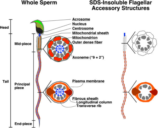
43 sperm cell diagram with labels
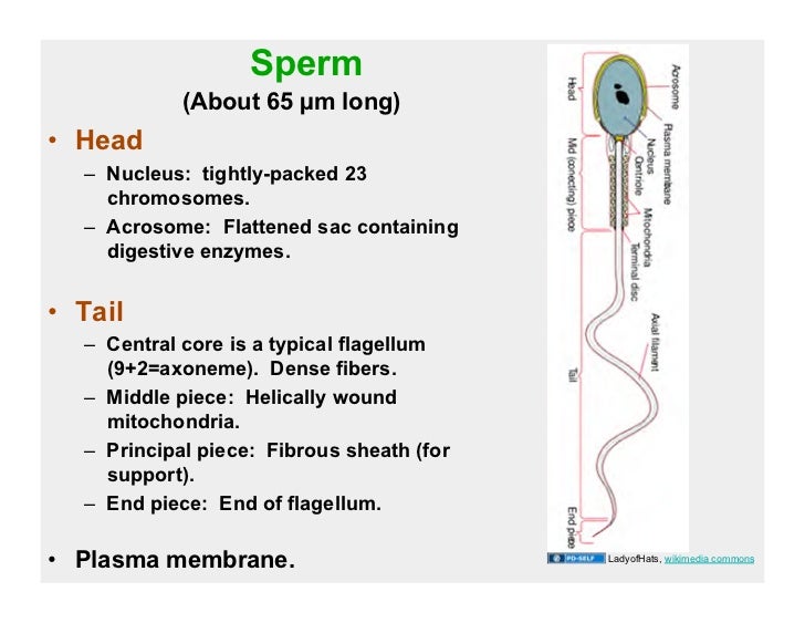
Sperm (Label) Diagram | Quizlet Start studying Sperm (Label). Learn vocabulary, terms, and more with flashcards, games, and other study tools. Home. Subjects. ... Gravity. Created by. Student_Nurse2020 PLUS. Terms in this set (12) Acrosome. A vesicle at the tip of a sperm cell that helps the sperm penetrate the egg. Plasma Membrane. A selectively-permeable phospholipid ... Sperm Cell, Egg Cell Diagram Label Worksheets (Differentiated) Three excellently differentiated worksheets. Engaging activity where pupils have to label the different parts of the male and femal gametes. Very well structured and scaffolded according to ability (from SEN to high ability). Excellent for visual learners. Compatible with all biology exam boards (including AQA, Edexcel, OCR).
quizlet.com › 440705001 › mastering-ch13-flash-cardsmastering ch.13 Flashcards & Practice Test | Quizlet Look carefully at the diagrams depicting different stages in meiosis in a cell where 2n = 6. Assume that the red chromosomes are of maternal origin and the blue chromosomes are of paternal origin. Drag the labels to fill in the targets beneath each diagram of a cell. Note that the diagrams are in no particular order.
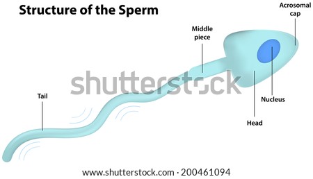
Sperm cell diagram with labels
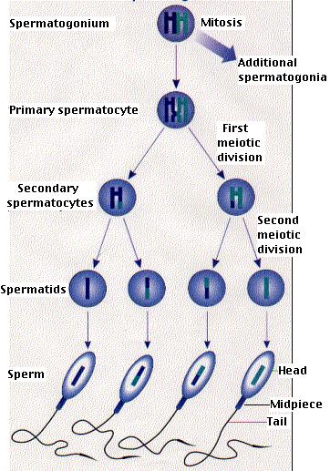
Week 16: Reproductive System Flashcards - Quizlet The acrosome is the structure at the tip of a sperm that contains enzymes to gain access to an egg during fertilization. Stem cells that give rise to sperm are spermatogonia. The middle embryonic germ layer that forms everything except the epidermis of the skin, nervous system, and epithelial linings and glands is the mesoderm. The Cell - ScienceQuiz.net The diagram shows a plant cell as seen under a microscope. Two of the labels are incorrect. What are they?? Vacuole and chloroplast? Vacuole and cytoplasm? Nucleus and chloroplast? Cell wall and cell membrane; The diagram shows a plant cell. ... the sperm cell? a red blood cell? a cheek cell? Testes: Anatomy and Function, Diagram, Conditions, and Health Tips The epididymis stores sperm cells until they're mature and ready for ejaculation. ... Explore the interactive 3-D diagram below to learn more about the testes. ... (2015). "Off-label" usage ...
Sperm cell diagram with labels. Sperm Cell Labeled Diagram Stock Vector (Royalty Free) 200461103 ... Find Sperm Cell Labeled Diagram stock images in HD and millions of other royalty-free stock photos, illustrations and vectors in the Shutterstock collection. Thousands of new, high-quality pictures added every day. A Labelled Diagram Of Meiosis with Detailed Explanation Meiosis is a type of cell division in which a single cell undergoes division twice to produce four haploid daughter cells. The cells produced are known as the sex cells or gametes (sperms and egg). The diagram of meiosis is beneficial for class 10 and 12 and is frequently asked in the examinations. The diagram of meiosis along with the ... Structure of Human Sperm: Check Types of Sperm - Embibe Explain the Structure of Human Sperm with Labelled Diagram Fig: Structure of a sperm cell Learn Exam Concepts on Embibe What is the Structure of Sperm? Human sperm is a microscopic structure whose shape is like a tadpole. It has flagella which make it motile. Its diameter is \ (2 - 5 {\rm { \mu m}},\) and its length is \ (60 {\rm { \mu m}}.\) Welcome to Butler County Recorders Office Copy and paste this code into your website. Your Link …
Draw a diagram of the microscopic structure of human sperm. Label the ... The above diagram is of the sperm cell. (a) Acrosome: It contains enzymes used for penetrating the female egg. (b) Nucleus: Contains the genetic material that the sperm has to pass on, a haploid genome because it contains only one copy of each chromosome. Structure and parts of a sperm cell - inviTRA This labelled diagram shows the structure of a sperm cell in detail, which has the following parts: Head With its spheric shape, it consists of a large nucleus, which at the same time contains an acrosome. The nucleus contains the genetic information and 23 chromosomes. It also secretes a hyaluronidase enzyme that destroys the hyaluronic acid ... An overview of sperm anatomy | Legacy Sperm is the male sex cell, also known as a gamete. Measuring approximately 0.05 millimeter (0.002 inch) long, sperm cells are made up of a few distinct parts: the tail, made up of protein fibers, which helps it "swim" toward the egg. the midpiece, or body, which contains mitochondria to power the sperm's movement. recorder.butlercountyohio.org › search_records › subdivisionWelcome to Butler County Recorders Office Copy and paste this code into your website. Your Link Name
Chloroplast - Wikipedia A chloroplast / ˈ k l ɔːr ə ˌ p l æ s t,-p l ɑː s t / is a type of membrane-bound organelle known as a plastid that conducts photosynthesis mostly in plant and algal cells.The photosynthetic pigment chlorophyll captures the energy from sunlight, converts it, and stores it in the energy-storage molecules ATP and NADPH while freeing oxygen from water in the cells. The ATP and NADPH … quizlet.com › 554874841 › week-16-reproductiveWeek 16: Reproductive System Flashcards - Quizlet The acrosome is the structure at the tip of a sperm that contains enzymes to gain access to an egg during fertilization. Stem cells that give rise to sperm are spermatogonia. The middle embryonic germ layer that forms everything except the epidermis of the skin, nervous system, and epithelial linings and glands is the mesoderm. Flow cytometry - Wikipedia Flow cytometry (FC) is a technique used to detect and measure physical and chemical characteristics of a population of cells or particles.. In this process, a sample containing cells or particles is suspended in a fluid and injected into the flow cytometer instrument. The sample is focused to ideally flow one cell at a time through a laser beam, where the light scattered is … What's the Function of a Sperm Cell? - Definition & Structure A spermatozoon, in plural spermatozoa, or sperm cell is the male reproductive cell that is expelled along with the seminal fluid or semen when a man ejaculates. In humans, spermatozoa determine the gender of the baby-to-be, which means that they can carry either the X or the Y chromosome.
Diagram and label sperm cell - Quizlet Only $2.99/month Diagram and label sperm cell STUDY Learn Flashcards Write Spell Test PLAY Match Gravity Created by Ike_SandersonTEACHER Terms in this set (4) Midsection of sperm contains mitochondria Sperm nucleus Contains haploid chromosomes Acrosome A vesicle at the tip of a sperm cell that helps the sperm penetrate the egg Flagellum
Diagram Of A Sperm Cell Illustrations, Royalty-Free Vector ... - iStock Browse 420 diagram of a sperm cell stock illustrations and vector graphics available royalty-free, or start a new search to explore more great stock images and vector art. Newest results structure of a sperm cell Education Chart of Biology for Reproduction Process of Human... Cell potency. From Totipotent to Pluripotent, Multipotent, and...
Draw the diagram of human sperm and label its parts. Write few lines ... Draw the diagram of human sperm and label its parts. Write few lines about it. Answer: The head area or acrosome contains enzymes which help to enter the egg during fertilization. Mitochondria acts as a power-house of the cell and there are several mitochondria available at the middle piece.
Draw the diagram of human sperm and label its parts. Write few lines ... Draw the diagram of human sperm and label its parts. Write few lines about it. Medium Solution Verified by Toppr The sperm cells are the haploid gametes which are produced in the male. There are different parts of the sperm cell. (a) Acrosome: This structure contains enzymes used for penetrating the female egg.
en.wikipedia.org › wiki › Lipid_bilayerLipid bilayer - Wikipedia When a vesicle is produced inside the cell and fuses with the plasma membrane to release its contents into the extracellular space, this process is known as exocytosis. In the reverse process, a region of the cell membrane will dimple inwards and eventually pinch off, enclosing a portion of the extracellular fluid to transport it into the cell.
6 Important Types of Membrane Proteins (With Diagram) Peripheral proteins are rich in amino acids with hydrophilic side chains that permit interaction with the surrounding water and with the polar surface of the lipid bilayer. Peripheral proteins on the cell’s exterior membrane surface often contain chains of sugars (i.e., they are glycoproteins). 2. Integral (Intrinsic) Proteins:
Draw and label the diagram of human sperm cell. - Sarthaks eConnect ... Draw and label the diagram of human sperm cell. reproduction; class-10; Share It On Facebook Twitter Email. 1 Answer +1 vote . answered Mar 2 by KalashAtagre (40.7k points) selected Mar 3 by KshitizKumar . Best answer. The diagram of human sperm cell ← Prev Question ...
mastering ch.13 Flashcards & Practice Test | Quizlet Look carefully at the diagrams depicting different stages in meiosis in a cell where 2n = 6. Assume that the red chromosomes are of maternal origin and the blue chromosomes are of paternal origin. Drag the labels to fill in the targets beneath each diagram of a cell. Note that the diagrams are in no particular order.
Lipid bilayer - Wikipedia The lipid bilayer is very thin compared to its lateral dimensions. If a typical mammalian cell (diameter ~10 micrometers) were magnified to the size of a watermelon (~1 ft/30 cm), the lipid bilayer making up the plasma membrane would be about as thick as a piece of office paper. Despite being only a few nanometers thick, the bilayer is composed of several distinct chemical …
labelled diagrams - the sperm cell labelled diagrams - the sperm cell to the right is a detailed 2D diagram of the sperm cell. there are many parts of a sperm cell. it is extremely small compared to the female egg.
Spermatogenesis Diagram & Function | What is the Process of Sperm ... Beneath the Sertoli cells are the spermatogonia, which are germ cells that will go through mitosis and ultimately create sperm. In humans, each day, roughly 25 million spermatogonia divide, and...
achieverstudent.comAchiever Student: Easy way to better grades. We write custom essay samples to help international students succeed with their studies Order your paper
How to draw Sperm Cell || Study of Human Spermatozoon diagram and label ... 'How to draw Sperm Cell || Study of Human Spermatozoon diagram and label the parts' is demonstrated in this video tutorial step by step.Sperm is the male rep...
› proteins › 6-important6 Important Types of Membrane Proteins (With Diagram) Peripheral proteins are rich in amino acids with hydrophilic side chains that permit interaction with the surrounding water and with the polar surface of the lipid bilayer. Peripheral proteins on the cell’s exterior membrane surface often contain chains of sugars (i.e., they are glycoproteins). 2. Integral (Intrinsic) Proteins:




Post a Comment for "43 sperm cell diagram with labels"